
Download
ORIGINAL ARTICLE
Therapeutic action of Kushen recipe extractive and its inhibitory effect on eotaxin in mouse models with contact dermatitis
Cheng Penga#, Quangang Zhub#, Jiyong Liuc##, Jishun Yangd##, Benming Youe###, Hualin Zhangf###, Yu Zhue, Jinhong Hue*
aDepartment of Health Management, Beidaihe Rehabilitation and Rest Center of PLA, Qinhuangdao, China
bDepartment of Pharmacy, Shanghai Skin Disease Hospital, Tongji University, Shanghai, China
cDepartment of Pharmacy, Fudan University Shanghai Cancer Center, Shanghai, China
dDepartment of Health Security Administration, Naval Medical Center, Naval Medical University, Shanghai, China
eDepartment of Pharmacy, The First Affiliated Hospital of PLA Naval Military Medical University, Shanghai, China
fDepartment of Pharmacy, Hospital of 81st Group Army PLA, Zhangjiakou, China
Abstract
Background: Treatment of skin allergic diseases remains a challenging research topic.
Objective: To investigate the effect of Kushen recipe extractive (KS) gel on contact dermatitis (CD) of mouse.
Methods: Allergic contact dermatitis (ACD) model of mouse was established. Immunohistochemical method (ICH) and flow cytometry method (FCM) were used to detect CD4+ and CD8+ T lymphocytes and explore the regulation effect of KS on the immune status of the organism. The expression status of eotaxin tissue was evaluated by real-time polymerase chain reaction (RT-PCR), ICH, and western blotting method. The survival rates of HaCaT cell and Fibroblasts affected by KS were detected by methyl thiazolyl tetrazolium (MTT) method. The inhibitory effect of KS on eotaxin produced by HaCaT cell and FBs induced by TNF-α and interleukin (IL)-4 were evaluated using RT-PCR and enzyme-linked immunosorbent assay methods. The inhibitory effect of KS on nuclear factor-κB (NF-κB) and Signal transducers and activators of transcription 6 (STAT6) activation induced by TNF-α and IL-4 was detected by electrophoretic mobility shift assay and western blotting methods.
Results: We confirmed that KS shows favorable therapeutic effect on CD, which can obviously inhibit eotaxin expression and Eosinophils recruitment in allergic skin of mouse, as well as regulate the immune status of the organism. Furthermore, KS and its main effective components can inhibit TNF-α and IL-4 induced upregulation of eotaxin via the two signal transduction pathways, NF-κB and STAT6.
Conclusions: The great importance of traditional Chinese recipe KS is evidenced by its therapeutic effect and mechanism in ACD of mouse.
Key words: eotaxin, allergy, skin, kushen recipe (KS), expression
*Corresponding author: Jinhong Hu, 168 Changhai Road, Yangpu District, Shanghai, 200433, China. Department of Pharmacy, The First Affiliated Hospital of PLA Naval Military Medical University, Shanghai 200433, China. Email address: [email protected]#First author; ##Second author; ###Third author
Received 7 November 2022; Accepted 8 February 2023; Available online 1 July 2023
Copyright: Peng C, et al.
License: This open access article is licensed under Creative Commons Attribution 4.0 International (CC BY 4.0). http://creativecommons.org/licenses/by/4.0/
Introduction
Traditional Chinese medicines show unique advantages in the treatment of allergic diseases, which can realize antianaphylaxis via multiple pathways and attach importance to the global immunologic balance. Their superior therapeutic action and less side effects have been well acknowledged, along with the therapeutic advantages and characteristics.1–3 Herein, finding new antiallergic drugs from traditional Chinese medicines has received increasing attention. In this regard, Kushen, an ancient recipe with simple formulation and reliable therapeutic effect, is anticipated to play its role. However, the traditional apozem restricts its further application and the ambiguous mechanism also raises difficulty in in-depth understanding and development. Modern high-tech methods are expected to better identify this traditional recipe and truly promote its potential applications and popularization.
Kushen recipe comprises two traditional Chinese medicines, Kushen and Schizonepeta, and it is noteworthy that the interactions among various components and changes of compatibility are important in revealing the mechanism of the recipe of traditional Chinese medicine. The transformation of components after compatibility constitutes the root in dialectical medication to treat diseases. Therefore, identifying the effective components and component changes after compatibility is quite important in disease prevention and treatment using traditional Chinese medicine, upon which the explanation on recipe can be thorough. Along with the efforts toward Kushen recipe (KS) analyses, one of its components Sophora flavescens Ait was found to show heat-clearing, damp-drying, wind-dispelling, parasite-killing, and itching relief effects, which can treat eczema, wet sore, itchy skin, leprosy, and scabies ringworm.4 However, it can be made into a recipe with Schizonepeta tenuifolia. The reasons for these findings are well explained using the theory of traditional Chinese medicine by the ancients, while modern scientific theory fails to reveal the mechanism. Some researchers believe that, compared with Oxymatrine (Omt), Matrine (Mat) shows superior effect in treating acute exudative inflammation and immunoregulation.5–8 In this regard, the compatibility between Sophora flavescens Ait and Schizonepeta tenuifolia converts part of oxidized alkaloids into reduced ones, which can potentially contribute to enhanced therapeutic effects. Meanwhile, the volatile oil in Schizonepeta tenuifolia also shows a favorable anti-inflammatory effect, which can serve as penetration enhancer when it is adopted for external use. The simple combination of the two medicines ensures the optimum therapeutic effect of the recipe.
Previous work has determined the material basis and compatibility of KS,9 upon which complete extracts of effective sites and extractive gels were obtained,10,11 paving a way for further experiments. In this work, based on the allergic contact dermatitis (ACD) model of mouse, the therapeutic effect of KS was evaluated and the mechanism of KS in treating allergic diseases was explored both in vivo and in vitro. The Kushen alkaloids can treat various Eosinophils (Eos)-related diseases, such as skin allergy, scar tissue proliferation, and liver fibrosis. Therefore, inhibiting Eos recruitment may serve as one of the treatment mechanisms. Bearing this in mind, we aim to explore its effect on the key chemotactic factor (eotaxin) in Eos recruitment.
Materials and Methods
Material basis and extraction method of Kushen recipe
Kushen recipe is composed of two traditional Chinese medicines, Kushen and Schizonepeta. Kushen belongs to the leguminous plant Sophora flavescens Ait, the main active components being alkaloids. Schizonepeta belongs to the dry overground part of the organum plant Schizonepeta tenuifolia, and the main active component is volatile oil. The mass ratio of Sophora flavescens Ait to Schizonepeta tenuifolia was 2:1 in the recipe. The alkaloid components and contents were analyzed by liquid chromatography (LC)/mass spectrum (MS), high performance liquid chromatography (HPLC), and potentiometric titration method, while the components and contents of volatile oil were analyzed using gas chromatogram (GC)/MS.9 It was found that alkaloid in Sophora flavescens Ait mainly contained four components: Mat, Omt, Sophocarpine (Sop), and Oxysophocarpine (Osp), which accounted for high proportion of >80% in the whole alkaloid, as shown in Figure S1. Specifically, Mat, Omt, Sop, and Osp accounted for 36.36 ± 0.86%, 18.88 ± 1.11%, 32.90 ± 3.16%, and 11.87 ± 1.37%, respectively. The main components of volatile oil in Schizonepeta tenuifolia were menthone, pulegone, and D-limonene. With the compatibility of the two medicines, the contents of effective components were obviously changed in comparison with single medicine. As shown in Figure S2, part of Mat converted into Omt and Sop converted into Osp. Similarly, the components of volatile oil also changed after compatibility, as shown in Figure S3.
Having ascertained the material basis of KS recipe, the extraction method of its effective site was designed to facilitate clinical applications. The main effective components were extracted completely to fabricate into a certain dosage form. Figures S4 and S5 show the extraction procedures and component analyses of the extractives.10,11
KS was then fabricated into gels that were suitable for percutaneous drug delivery, which was then subjected to in vitro transdermal experiment using Spragne–Dawley (SD) rat. The aqueous extractive and purified extractives (and gels) were compared, which showed that transdermal effect can be greatly enhanced after the main components of KS were subjected to purified extraction and fabricated into gels,12 as shown in Figure S6.
Therapeutic action and mechanism of KS in ACD mouse
For therapeutic action observation and mechanism of KS on ACD mouse, 2,4-Dinitrofluorobenzene (DNFB) was smeared onto mouse ear six times before and after activation to ensure a stable drug effect. KS with two different concentrations (1 and 5%) constituted the therapeutic groups. Dexamethasone sodium phosphate (Dex) and Cetirizine Hydrochloride (Cet) were set as positive control groups. The ear thickness of mouse was measured to evaluate the effect of drug therapy. The pathological section observation after hematoxylin and eosin (HE) staining was utilized to verify the drug improvement effect on the infiltration of inflammatory cells.
Flow cytometery (FCM) and immunohistochemical (ICH) methods were adopted to detect the CD4+ and CD8+ T-lymphocytes in blood and tissue, respectively. They were designed to investigate the regulation effect of KS on the immune status of organism. Real-time polymerase chain reaction (RT-PCR), western blotting, and ICH methods were used to detect the expression status of eotaxin (8.4 kD) tissue in ACD mouse model.
The nuclear translocation effects of KS and its main components, Mat, Omt, Sop, Osp, and schizonepetae volatile oil (SVO), on nuclear factor-κB (NF-κB) induced by TNF-α were investigated. Specifically, the drugs were co-incubated with the cells for 24 h, followed by TNF-α stimulation on HaCaT cells and Fibroblast (FBs) for 45 and 60 min, respectively. The effects of KS and its main effective components on pSTAT6 (100kD) were also investigated. Typically, the drugs were co-cubated with cells for 24 h, and both the HaCaT cells and FBs were stimulated by Interleukin (IL)-4 for 30 min. KS, Mat, Omt, Sop, Osp, SVO, Dex, and Cet with different concentrations were individually interacted with HaCaT cells and FBs for 24 h, which were then co-cubated with TNF-α (100 ng/mL) or IL-4 (10 ng/mL) for 12 h to detect the expression. It should be noted that the HaCaT cell line and 6th to 10th generation FBs were adopted in this work.
Inhibitory mechanism of KS on eotaxin expression in skin cells
The methyl thiazolyl tetrazolium (MTT) method was adopted to evaluate the effects of KS, SVO, Mat, Omt, Sop, Osp, Cet, and Dex on the survival rates of HaCaT cells and FBs within 36 h. RT-PCR and enzyme-linked immunosorbent assay (ELISA) methods were used to detect the inhibitory effects of KS, SVO, Mat, Omt, Sop, and Osp on eotaxin produced by HaCaT cells and FBs, which were induced by TNF-α and IL-4, respectively. Meanwhile, Cet and Dex were taken as control experiments and used to intervene in the process. Electrophoretic mobility shift assay (EMSA) and western blotting methods were individually utilized to evaluate the inhibitory effects of Mat, Omt, Sop, Osp, and SVO on the activation of NF-κB and Signal transducers and activators of transcription 6 (STAT6), which were induced by TNF-α and IL-4, respectively.
Results and Analysis
Therapeutic action and mechanism of KS on mouse ACD
Therapeutic effect observation of KS on ACD mouse
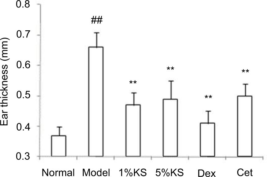
After treating with 1 and 5% KS, the swelling of mouse ear decreased, and no obvious adverse reaction was observed. The measurement of mouse ear thickness indicates that KS treatment can significantly decrease the swelling (P < 0.01), but there is no difference between the 1 and 5% KS treatment groups. Specifically, the ear thickness in control group, model group, and 1 and 5% KS treatment groups are 0.37 ± 0.03 mm, 0.66 ± 0.05 mm, 0.47 ± 0.04 mm, and 0.49 ± 0.06 mm, respectively. Compared with the Cet (0.50 ± 0.04 mm) group, there is no significant difference in the two KS treatment groups, while the therapeutic effect is inferior than that of the Dex group (0.41 ± 0.04 mm), as shown in Figure 1.
Figure 1 The inhibitory effect of Kushen recipe (KS) on ear swelling in mouse allergic contact dermatitis (ACD) model. (n = 12) ##p < 0.01, vs. normal group; **p < 0.01, vs. model group.
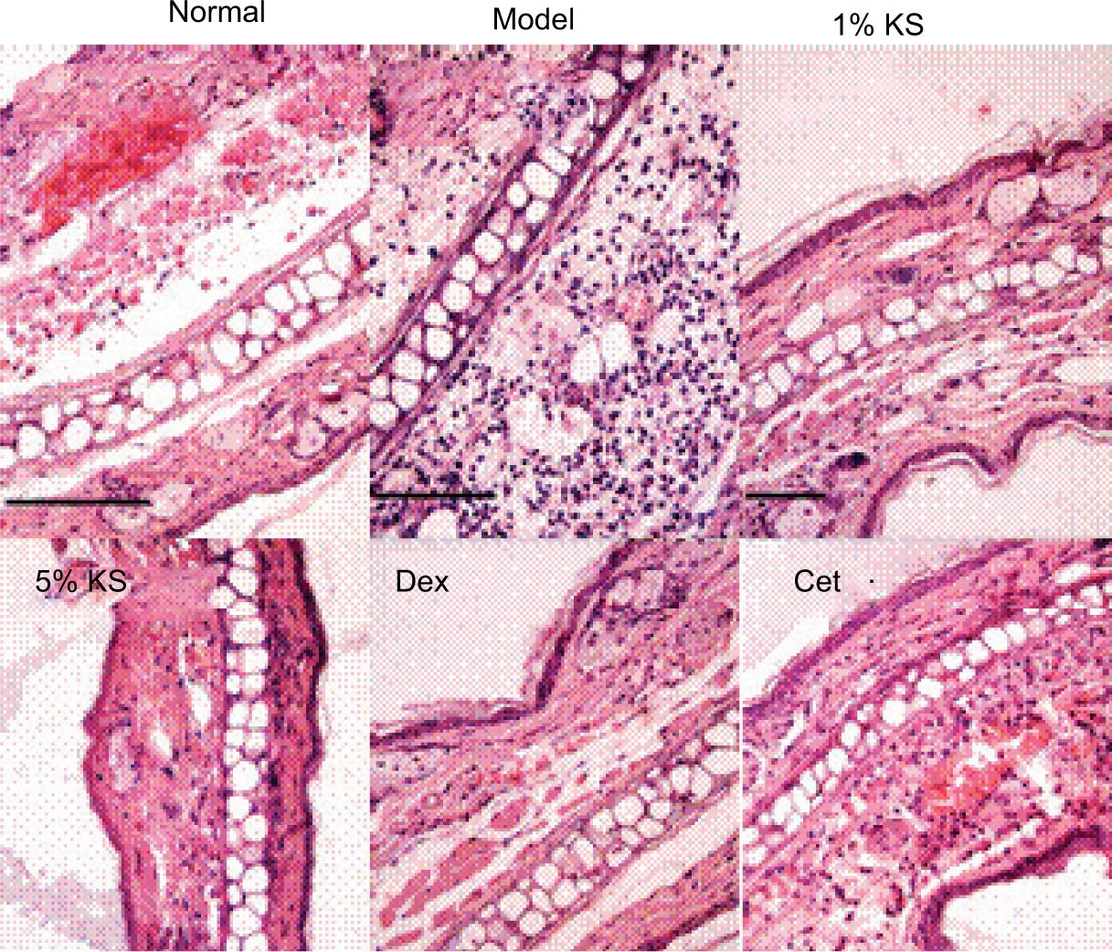
According to the H-E staining results, KS can significantly improve the infiltration degree of inflammatory cells in the skin tissue of mouse ACD model. The positive control groups, Dex and Cet, also indicate a favorable therapeutic effect, as shown in Figure 2.
Figure 2 Effect of Kushen recipe (KS) on pathological changes in mouse allergic contact dermatitis (ACD) model. The scale bars are 100 µm.
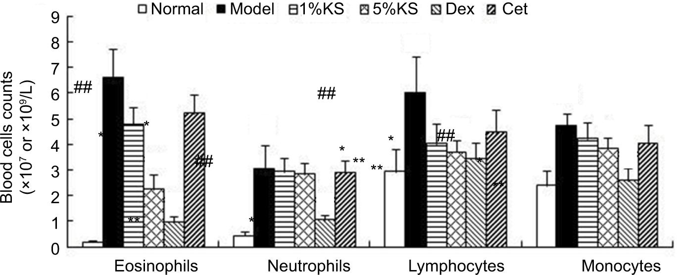
Also, 1 and 5% KS can obviously lower the Eos counting in mouse ACD blood (P < 0.05 and P < 0.01 for 1 and 5% KS, respectively). Although it readily lowers Lym (P < 0.05), the regulation over neutrophils (Neu) and monocytes (Mon) was not so obvious. Compared with positive control drug, KS and Cet have similar effect on hemocyte. Higher effect degree is found in Dex, which shows intense inhibitory effect in terms of the recruitment of four leukocytes in blood, as shown in Figure 3.
Figure 3 Effect on blood cell counts of Kushen recipe (KS) in mouse allergic contact dermatitis (ACD) model (n = 6). Eosinophils (Eos) (×107/L), Lym (×109/L), Neutrophils (Neu) (×109/L), monocytes (Mon) (×107/L). ## p < 0.01, vs. normal group, * p < 0.05, vs. model group, ** p < 0.01, vs. model group.
Effect of KS on the CD4+ and CD8+ T cells in the skin tissue and blood of mouse ACD
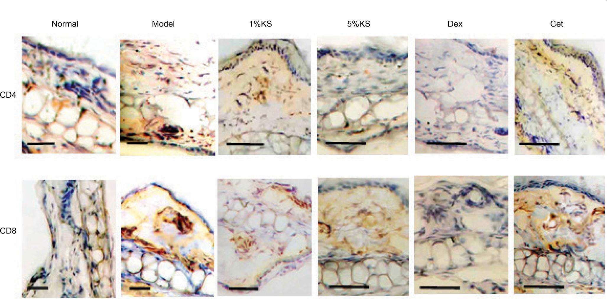
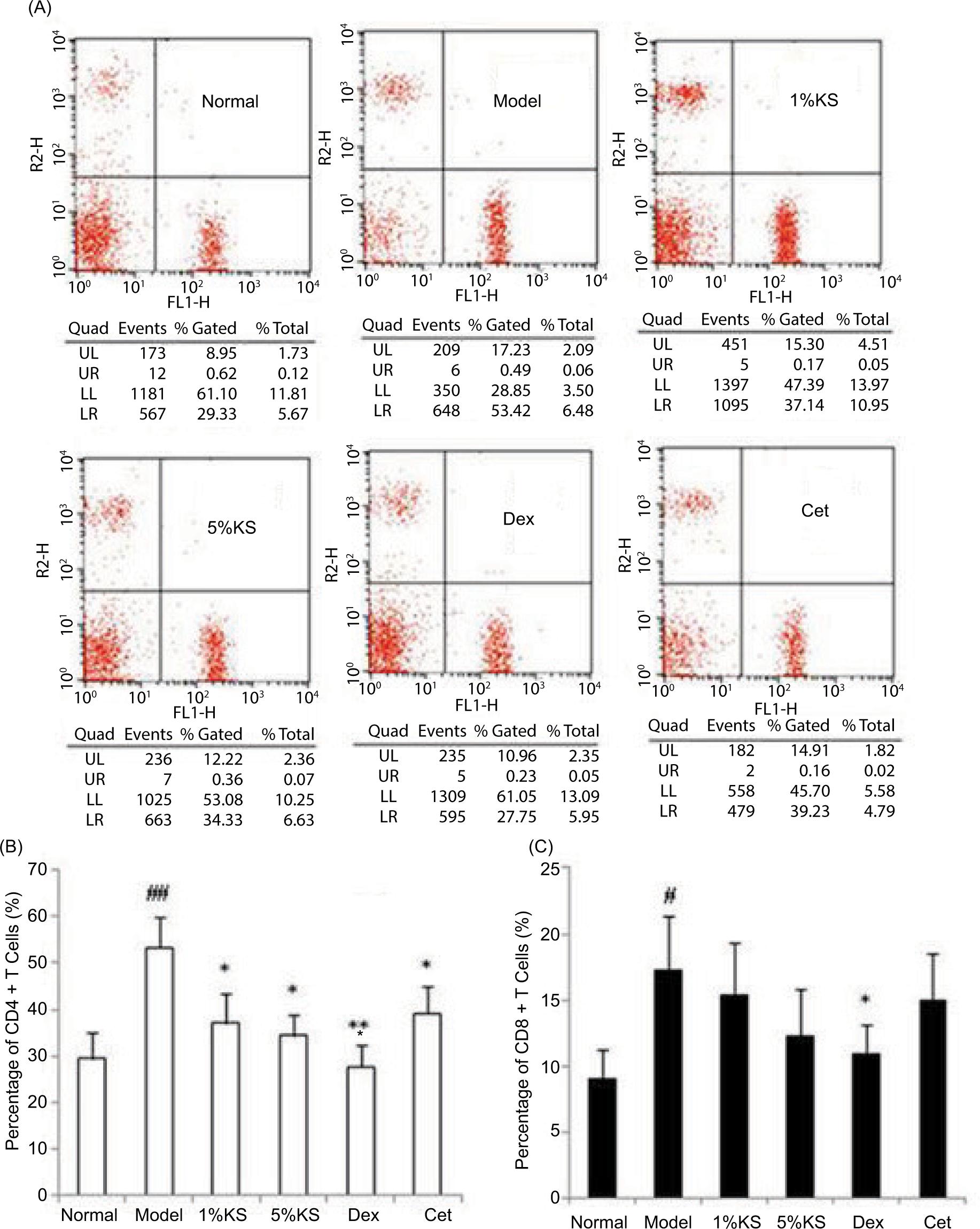
The ICH staining results of the tissue reveal that KS can, to some extent, inhibit the infiltration of CD4+ and CD8+ T cells of the mice induced by DNFB. Cet also shows this inhibitory effect, which turns to be more evident for Dex, as shown in Figure 4. In addition, KS, Cet, and Dex show inhibitory effect on CD4+ T cell in mouse blood, while only 5% KS and Dex show inhibitory effect on CD8+ T cell with statistic difference (P < 0.05), as displayed in Figure 5.
Figure 4 Effect of Kushen recipe (KS) on CD4+ and CD8+ T cells infiltration in skin of mouse allergic contact dermatitis (ACD). The scale bars are 100 µm.
Figure 5 Effect of Kushen recipe (KS) on CD4+ and CD8+ T cells in the blood of mouse allergic contact dermatitis (ACD) model. (n = 6) A: Scatterplot of flow cytometry, B: Percentage of CD4+ T cells, C: Percentage of CD8+ T cells. #p < 0.05, vs. normal group, ## p < 0.01, vs. normal group, * p < 0.05, vs. model group, ** p < 0.01, vs. model group.
Effect of KS on eotaxin mRNA expression in skin tissue of mouse ACD
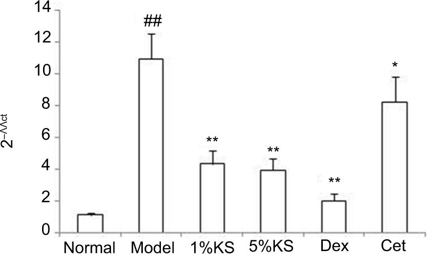
The 1 and 5% KS can significantly inhibit the expression of eotaxin messenger ribonucleic acid (mRNA) in skin tissue of mouse ACD (P < 0.01), while the two groups indicate no significant difference. Also, Dex can significantly inhibit the expression of eotaxin mRNA (P < 0.01), showing more favorable effect than that of KS. Cet can, to some extent, inhibit the expression of eotaxin mRNA, while the effect is inferior than that of KS and Dex, as shown in Figure 6.
Figure 6 The inhibitory effect of Kushen recipe (KS) on eotaxin messenger ribonucleic acid (mRNA) in the skin of mouse allergic contact dermatitis (ACD) model (n = 6). ## p < 0.01, vs. normal group, * p < 0.05, vs. model group, ** p < 0.01, vs. model group.
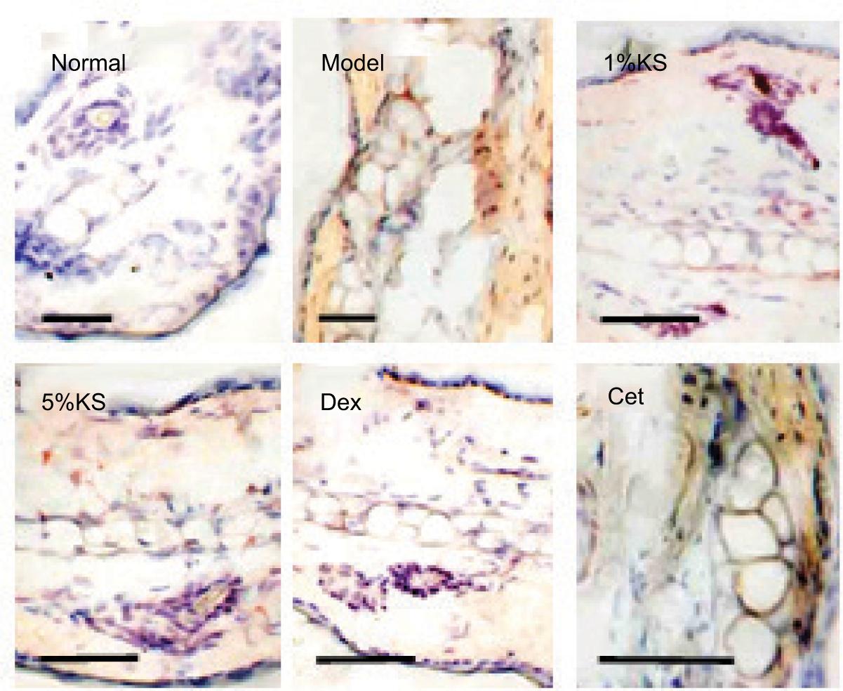
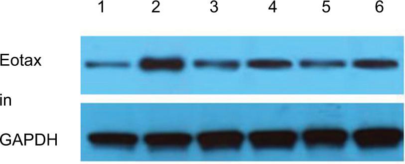
The ICH results indicate that KS (1 and 5%), Cet, and Dex can improve the secretion of eotaxin in the tissue, but Cet shows poorer inhibitory effect on eotaxin, as indicated in Figure 7. It is also found that both 1 and 5% KS can inhibit eotaxin secretion; such an effect appears to be more and less evident in Dex and Cet, respectively The internal reference protein is Glyceraldehyde phosphate dehydrogenase (GAPDH) (36 kD), as shown in Figure 8.
Figure 7 Inhibitory effect of Kushen recipe (KS) on eotaxin secretion in the skin of mouse allergic contact dermatitis (ACD) detected by immunohistochemistry. The scale bars are 100 µm.
Figure 8 The inhibitory effect of Kushen recipe (KS) on eotaxin secretion in the skin of mouse allergic contact dermatitis (ACD) model. 1: Normal group, 2: Model group, 3: 1% KS, 4: 5% KS, 5: Cetirizine Hydrochloride (Dex), 6: Cetirizine Hydrochloride (Cet).
Inhibitory mechanism of KS on eotaxin expression in skin cells
Effect of drugs on the survival rates of HaCaT cells and FBs
The MTT experimental results reveal that the survival rates were not significantly affected (≥90%) when the concentrations of KS and SVO were ≤500 μg/mL; Mat, Omt, Sop, and Osp were ≤1000 μM; Cet was ≤200 μM; and Dex was ≤100 μM. In this situation, there was also no stimulated fission and proliferation.
Effect of KS on the upregulation of eotaxin mRNA in HaCaT cell and FBs induced by TNF-α and IL-4
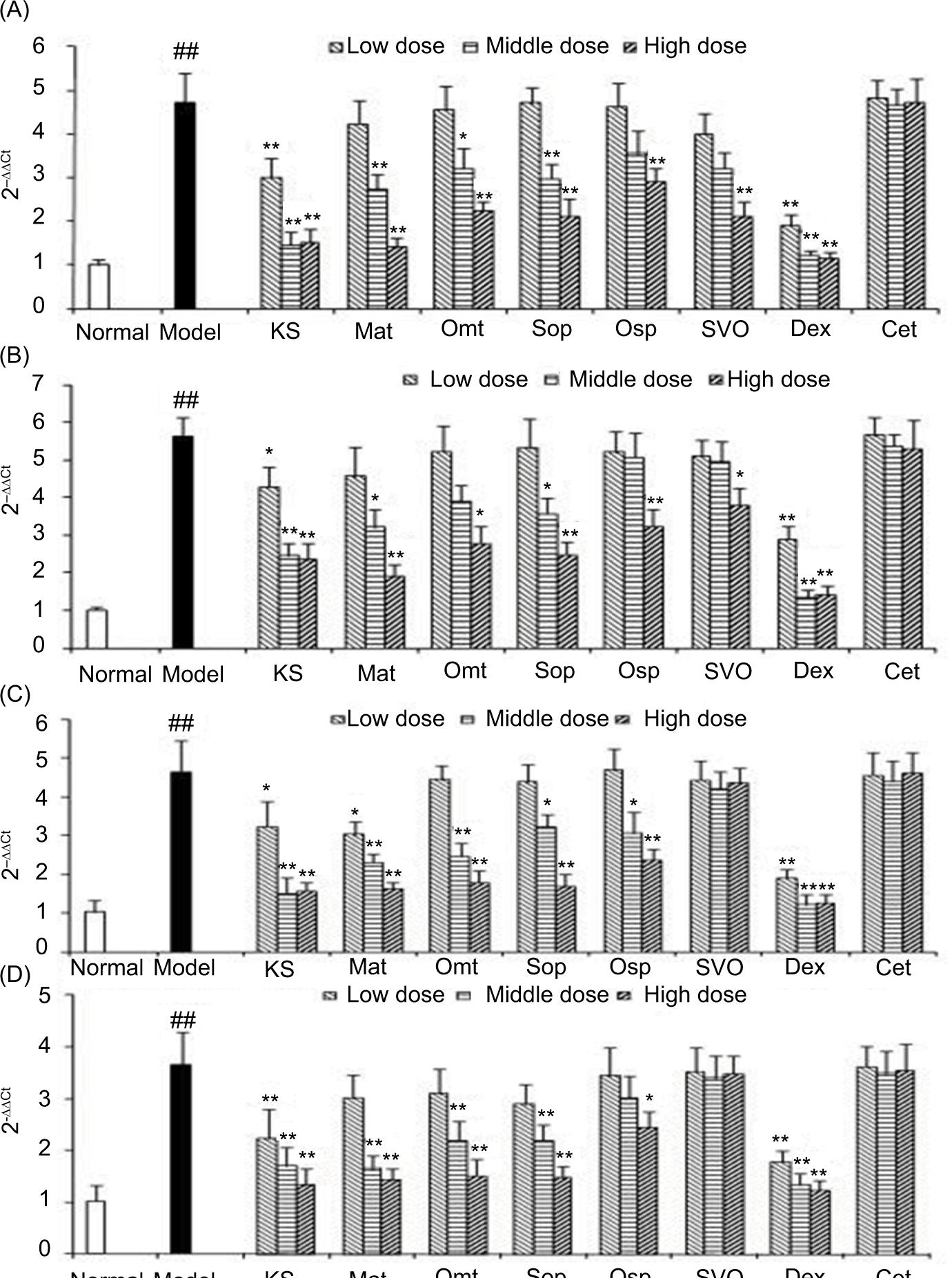
As displayed in Figures 9A–9D, similar tendency can be found in the two cells (HaCaT cells and FBs). KS and its internal components show an inhibitory effect on the expression of eotaxin mRNA, which are basically dose dependent. Among these, Mat presents the highest inhibitory effect, while SVO only exhibits an inhibitory effect toward eotaxin upregulation induced by TNF-α. Also, SVO shows no obvious effect on the regulation of eotaxin induced by IL-4. It is also found that Dex shows obvious and intense inhibitory effect on eotaxin mRNA, but this was not identified in Cet.
Figure 9 Drugs action on eotaxin messenger ribonucleic acid (mRNA) expression induced by TNF-α in HaCaT cells (A) and Fibroblast (FBs) (B); Drugs action on eotaxin mRNA expression induced by IL-4 in HaCaT cells (C) and FBs (D). (n = 6). ## p < 0.01, vs. normal group, *p < 0.05, vs. model group, **p < 0.01, vs. model group.
Effect of KS on eotaxin secretion in HaCaT cells and FBs induced by TNF-α and IL-4
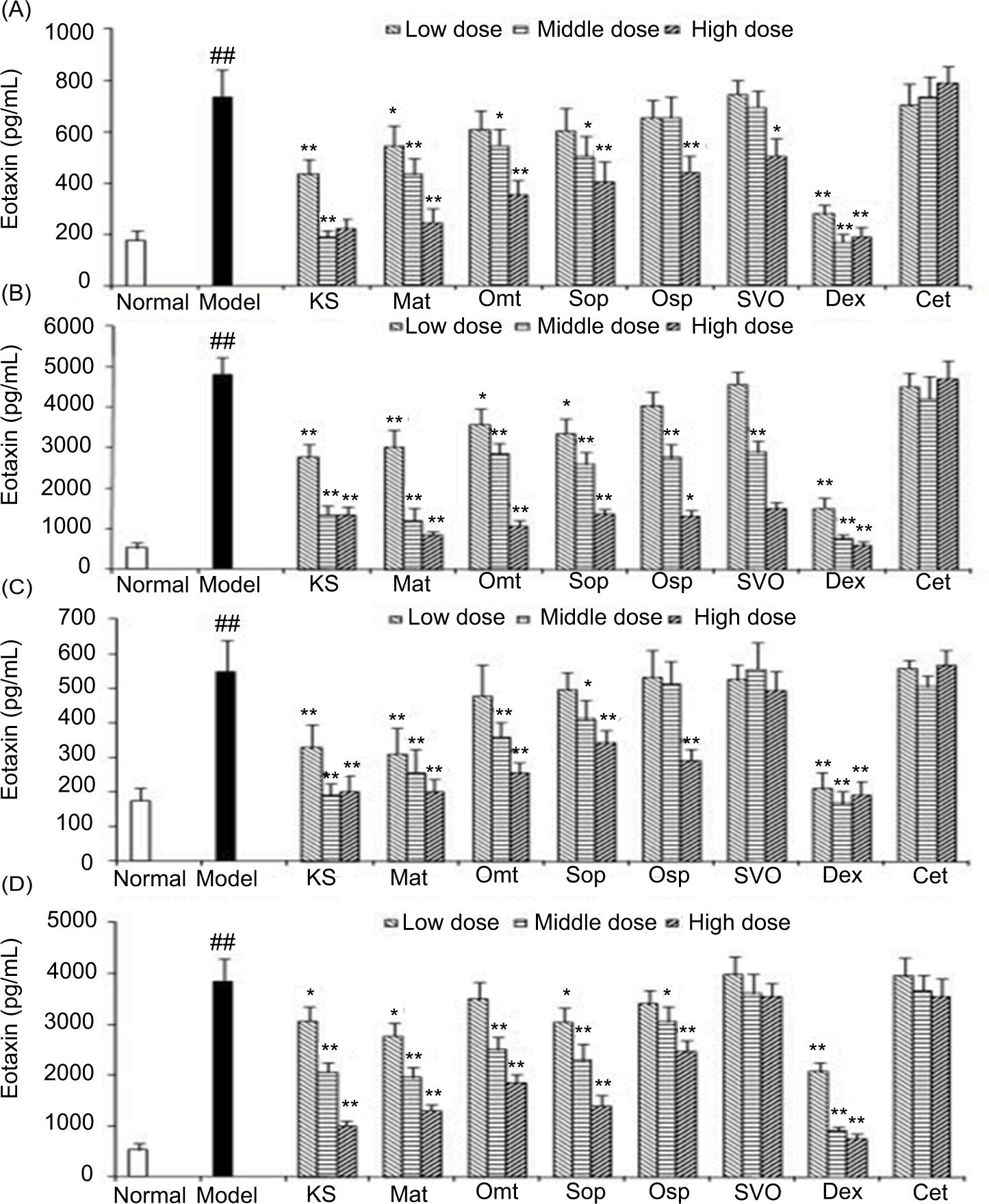
The secretion status of eotaxin resembles that of mRNA expression. Specifically, Cet exhibits no obvious inhibitory effect toward eotaxin secretion of the two cells induced by TNF-α (100 ng/mL) or IL-4 (10 ng/mL), SVO only shows certain inhibitory effect toward eotaxin release induced by TNF-α. Other components present different inhibitory effects toward eotaxin secretion induced by TNF-α or IL-4, which were basically dose dependent. Among these, Dex exhibits stronger inhibitory effect. The inhibitory effect of alkaloids in KS and volatile oil comes with a sequence of KS>Mat>Omt≈Sop>Osp≈SVO, as displayed in Figures 10A–D.
Figure 10 Drugs action on eotaxin secretion induced by TNF-α in HaCaT cells (A) and Fibroblast (FBs) (B); Drugs action on eotaxin secretion induced by IL-4 in HaCaT cells (C) and FBs (D). (n = 6) ## p < 0.01, vs. normal group, *p < 0.05, vs. model group, **p < 0.01, vs. model group.
In addition, the effects of above-described drugs on the basic expression of eotaxin were also evaluated; the results reveal that only Dex shows certain inhibitory effect on the basic expression of eotaxin.
Transcription mechanism of the inhibitory effect of KS on eotaxin in HaCaT cells and FBs
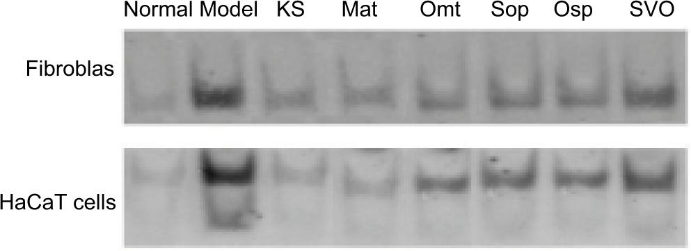
Effect of KS on the nuclear translocation of NF-κB induced by TNF-α
As shown in Figure 11, TNF-α can obviously induce the nuclear translocation of NF-κB, while KS and its components can inhibit the intranuclear transfer of NF-κB to some extent. These findings suggest that it may be one of the main mechanisms that inhibit eotaxin expression induced by TNF-α.
Figure 11 The action of Kushen recipe (KS) and its components on nuclear factor-κB (NF-κB) nuclear translocation.
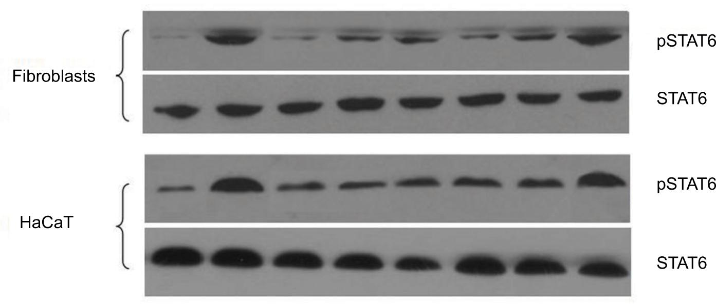
Effect of KS on STAT6 phosphorylation induced by IL-4
STAT6 (94 kD) phosphorylation induced by IL-4 is also one of the mechanisms for eotaxin production. In this regard, the effects of KS and its main effective components on pSTAT6 (100 kD) were investigated, as shown in Figure 12. Drugs and cells were co-incubated for 24 h, then IL-4 was utilized to stimulate HaCaT cells and FBs for 30 min. Except SVO, all other drug components are found to show inhibitory effects on STAT6 phosphorylation.
Figure 12 The action of Kushen recipe (KS) and its components on STAT6 phosphorylation.
Discussion
In this work, the complete extraction methods of the effective sites in the recipe were designed and fabricated into gel, upon which the transdermal effect was verified. Along with the established procedures, the appropriate carrier and simple method for potential application were found. The therapeutic effect of KS gel on mouse ACD was investigated. The results show that KS can significantly improve the swelling degree induced by the allergic inflammation of mouse ear skin and the infiltration degree of skin inflammatory cells, which can also decrease the number of inflammatory cells in blood (including Eos), showing immune regulation effect upon affecting CD4+ and CD8+ T cells. The mechanism study shows that KS can inhibit the expression amount of eotaxin in the skin of mouse ACD, the key chemotactic factor of Eos. Accordingly, it prohibits the tissue infiltration of Eos (the leading effector cell in atopic dermatitis [AD]) and blocks the occurrence of inflammation as well as alleviates tissue damage. The combined results suggest that eotaxin is the main therapeutic target in KS-treated AD. At the cellular level, the inhibitory effect of KS on eotaxin was realized via NF-κB and STAT6 signal pathways. To sum up, modern technologies were utilized to investigate and explore the material basis and working principle of traditional recipe KS, which may pave a way for its further development and application.
There remains some issues to be discussed. The dose for animals is based on the dose for humans and converted according to the species, while percutaneous drug delivery is of particularity. To date, no unambiguous references have been proposed to convert the dose based on human dose, while the commercially available species were adopted for the positive control Dex group. Therefore, two dose groups (1 and 5%) were designed for KS based on human dose (the gel content in commercially available Omt is 1%). The two groups show no significant difference in terms of the ability in inhibiting eotaxin production, while 5% KS group shows superior ability to inhibit Eos in blood than that of 1% KS. For percutaneous drug delivery under a certain dose, the drug accumulated in skin reaches saturation when drug permeates through skin, upon which the difference in external drug concentration differs from subcutaneous drug dose. Therefore, the dose-dependent inhibitory effect of KS toward eotaxin in cell experiment was not reflected in the in vivo experiment. However, the concentration difference of drug after entering into blood via transdermic absorption occurred, that is, the drug with higher concentration entered into blood with higher amount and stronger effect. Furthermore, the amount of Eos in blood is regulated by various factors including IL-5, which is not directly correlated with the secretion amount of eotaxin in tissue. The combined findings reveal that the pharmacologic effects and targets of KS were not single ones, which may regulate the immune status of an organism via multiple ways.
Kushen alkaloids have been verified to possess multiple pharmacological activities and low rate of adverse reactions, which have already been fabricated into various dosage forms in clinic (injection, oral medication, external use, etc.) to treat liver cirrhosis, arrhythmia, and inflammatory diseases.13,14 They can inhibit or downregulate the expression of TGF-β, TNF-α, Fas, and FasL, as well as inhibit some biological functions including the activity of protein kinase C.15,16 Other reports also claimed their immune regulation effect on an organism,17 which also shows obvious inhibitory effect on the cellular immunity of immunocompromised mice and strengthens its nonspecific immunity.18 In this work, we found that eotaxin is a new target for KS and its main active components, which can also regulate the immune status of organism by inhibiting CD4+ T cell, thus inhibiting inflammation.
In the cell experiment, the effects of four alkaloids of the main active components (Mat, Omt, Sop, and Osp) and SVO on eotaxin were also compared; the results established that the inhibitory effect of KS on eotaxin was realized via the above-listed components. Also, the contribution of these components in inhibiting eotaxin was also compared; it was found that the four alkaloids with the same mother-ring show inhibitory effect on eotaxin, while the subtle structural difference led to different strength of pharmacological activity. It is envisioned that the mother-ring can be modified to synthesize and screen compounds with stronger pharmacological activity, upon which new antiallergic drugs can be developed.
In addition, we also found that SVO can partly inhibit the upregulation of eotaxin induced by TNF-α, which however has no effect on eotaxin secretion induced by IL-4. Further experiment reveals that SVO shows no obvious effect on IL-4-STAT6 path, while other alkaloids can affect the production of eotaxin via TNF-α-NF-κB and IL-4-STAT6 paths. Except for Dex, other drugs show no effect on the basic expression of eotaxin in cell. It was then speculated that the mechanism of the basic expression of eotaxin differs from that induced by TNF-α and IL-4, the mechanism in more detail remains to be explored.
Two drugs were adopted as positive control groups in an animal experiment, the intense immune-inhibitory effect of Dex was verified, and its inhibitory effect on eotaxin has been reported.19,20 As an antagonist of H2 acceptor, Cet is widely used for the treatment of allergic diseases but its inhibitory effect on eotaxin remains ambiguous. Previous report claimed that it can inhibit the production of eotaxin in mouse abdominal inflammation.21 In this work, Cet shows inhibitory effect on eotaxin production in ACD mouse ear and decreases Eos amount in blood. However, in cell experiments, Cet shows no obvious effect on eotaxin upregulation induced by inflammatory factors. It is believed that the mechanism of AD in vivo involves multiple factors, and the inhibitory effect of Cet on eotaxin may be caused by indirect factors other than direct ones. As for the cell experiment in relatively simple environment, it was found that Cet cannot directly inhibit eotaxin secretion induced by inflammatory factors.
Although KS gel is generally adopted for external use in treating skin AD, our work proves that it can also play a role by directly inhibiting eotaxin production by skin cells after the transdermal process. Its inhibitory effect on eotaxin constitutes the key procedure, but this does not mean that KS only plays a role in this single procedure. The key point lies in the fact that KS can play a general role when locally working on the key position, which can be reflected by its effect on inflammatory granulocytes including Eos in blood and CD4+ T cells. Our previous work focused on the in vivo pharmacokinetics of transdermal drug delivery of Omt gel; the concentration of Omt and its metabolite Mat in subcutaneous tissue and blood were detected by microdialysis technique.22 Other researchers also carried out the bioavailability study of Mat cream.23 All these results found that Kushen alkaloids can reach up to effective plasma concentration and further play a general role after transdermal drug delivery.
It is summarized that the pathogenesis of allergic diseases is complicated, the therapeutic mechanisms using traditional Chinese recipe also link with each other, which shall block the key procedures of inflammation occurrence and regulate the overall immune balance. Local effect and general effect are closely coupled to realize a therapeutic effect.
Conclusion
In this work, the therapeutic action of KS gel on mouse ACD and its capability in immune regulation was verified. The combined findings suggest that KS recipe shows upregulation effect on eotaxin in mouse ACD, which verifies if eotaxin can serve as a new target for the pharmacologic action of KS and its main component, that is, Kushen alkaloids. Furthermore, it was verified whether KS recipe extractive and its main components, which inhibited eotaxin secretion induced by TNF-α and IL-4, were realized by two transcription pathways, NF-κB and STAT6.
REFERENCES
1. Huang SK, Lai CS, Chang YS, et al. Utilization pattern and drug use of traditional Chinese medicine, western medicine, and integrated Chinese-western medicine treatments for allergic rhinitis under the national health insurance program in Taiwan. J Altern Complement Med. 2016;22(10):832–840. 10.1089/acm.2015.0080
2. Lin PY, Chu CH, Chang FY, et al. Trends and prescription patterns of traditional Chinese medicine use among subjects with allergic diseases: a nationwide population-based study. World Allergy Organ J. 2019;12(2):100001. 10.1016/j.waojou.2018.11.001
3. Shao YY, Zhou YM, Hu M, et al. The anti-allergic rhinitis effect of traditional Chinese medicine of Shenqi by regulating mast cell degranulation and Th1/Th2 cytokine balance. Molecules. 2017;22(3):504. 10.3390/molecules22030504
4. Zhang W, Liu X, Fan H, et al. Separation and purification of alkaloids from Sophora flavescens Ait. by focused microwave-assisted aqueous two-phase extraction coupled with reversed micellar extraction. Indust Crops Prod. 2016;86:231–238. 10.1016/j.indcrop.2016.03.052
5. Liou CJ, Lai YR, Chen YL, et al. Matrine attenuates COX-2 and ICAM-1 expressions in human lung epithelial cells and prevents acute lung injury in LPS-induced mice. Mediators Inflamm. 2016;2016:3630485. 10.1155/2016/3630485
6. Wu C, Xu Z, Gai RH, et al. Matrine ameliorates spontaneously developed colitis in interleukin-10-deficient mice. Int Immunopharmacol. 2016;36:256–262. 10.1016/j.intimp.2016.04.038
7. Wang H. Effect of matrine injection on chemosensitization and quality of life and immune function of patients with cervical cancer. Basic Pharmacol Toxicol. 2019;125:20–21.
8. Sun N, Sun PP, Lv HP, et al. Matrine displayed antiviral activity in porcine alveolar macrophages co-infected by porcine reproductive and respiratory syndrome virus and porcine circovirus type 2. Sci Rep. 2016;6:24401. 10.1038/srep24401
9. Peng C, Hu JH, Zhu QG, et al. Component analysis and compatibility mechanism of the effective site of Kushe recipe. Chin Trad Herb Drugs. 2008;39(11):32–36. (in Chinese)
10. Peng C, Hu JH, Zhu QG, et al. Selection of optimum extraction technology for Radix Sophorae Flavescentis in Kushen recipe with orthogonal test. Pharm Care Res. 2007;4(2):124–127. (in Chinese)
11. Peng C, Hu JH, Zhu QG, et al. Technical study on the effective site of Kushen recipe extracted by alcohol-extraction-macroporous resin purification method. Chin Trad Patent Med. 2008;30(7):989–993. (in Chinese)
12. Peng C, Hu JH, Zhu QG, et al. Studies on cutaneous permeation in vitro of Kushen recipe gel. Chin J Chin Mater Med. 2007;32(18):1870–1874. (in Chinese)
13. Rashid HU, Xu YM, Muhammad Y, et al. Research advances on anticancer activities of matrine and its derivatives: an updated overview. Eur J Med Chem. 2019;161:205–238. 10.1016/j.ejmech.2018.10.037
14. Li Y, Wang G, Liu J, et al. Quinolizidine alkaloids derivatives from Sophora alopecuroides Linn: bioactivities, structure-activity relationships and preliminary molecular mechanisms. Eur J Med Chem. 2020;188:111972. 10.1016/j.ejmech.2019.111972
15. Huang JL, Xu H. Matrine: bioactivities and structural modifications. Curr Opin Med Chem. 2016;16(28):3365–3378. 10.2174/1568026616666160506131012
16. Jiang JS, Wang GJ. Matrine protects PC12 cells from lipopolysaccharide-evoked inflammatory injury via upregulation of miR-9. Pharmaceut Biol. 2020;58(1):314–320. 10.1080/13880209.2020.1719165
17. Nie JJ, Chen F, Wang FW, et al. Pharmacokinetics and bioavailability of matrine in rats by UPLC-MS/MS. Latin Am J Pharm. 2020;39(3):612–616.
18. Li NF, Zhao JX, Di TT, et al. Matrine alleviates imiquimod-induced psoriasiform dermatitis in BALB/c mice via dendritic cell regulation. Int J Clin Exp Pathol. 2018;11(11):5232–5240.
19. Lv YX, Dai MY, Wang MZ, et al. Anti-inflammatory property of galectin-1 in a murine model of allergic airway inflammation. J Immunol Res. 2019;2019:9705327. 10.1155/2019/9705327
20. Correa MP, Andrade FEC, Gimenes AD, et al. Anti-inflammatory effect of galectin-1 in a murine model of atopic dermatitis. J Mol Med. 2017;95(9):1005–1015. 10.1007/s00109-017-1566-9
21. Shimizu T, Nishihira J, Watanabe H, Abe R, Ishibashi T, Shimizu H. Cetirizine, an H1-receptor antagonist, suppresses the expression of macrophage migration inhibitory factor: its potential anti-inflammatory action. Clin Exp Allergy. 2004;34(1):103–109. 10.1111/j.1365-2222.2004.01836.x
22. Zheng H, Chen G, Shi L, et al. Determination of oxymatrine and its metabolite matrine in rat blood and dermal microdialysates by high throughput liquid chromatography/tandem mass spectrometry. J Pharm Biomed Anal. 2009;49(2):427–433. 10.1016/j.jpba.2008.11.032
23. Wei H, Li ZW, Wang Q, et al. Studies on pharmacolinetics and bioavailability of matrine creams. Chin Pharm J. 2001;36(3):183–185. (in Chinese)
Supplementary
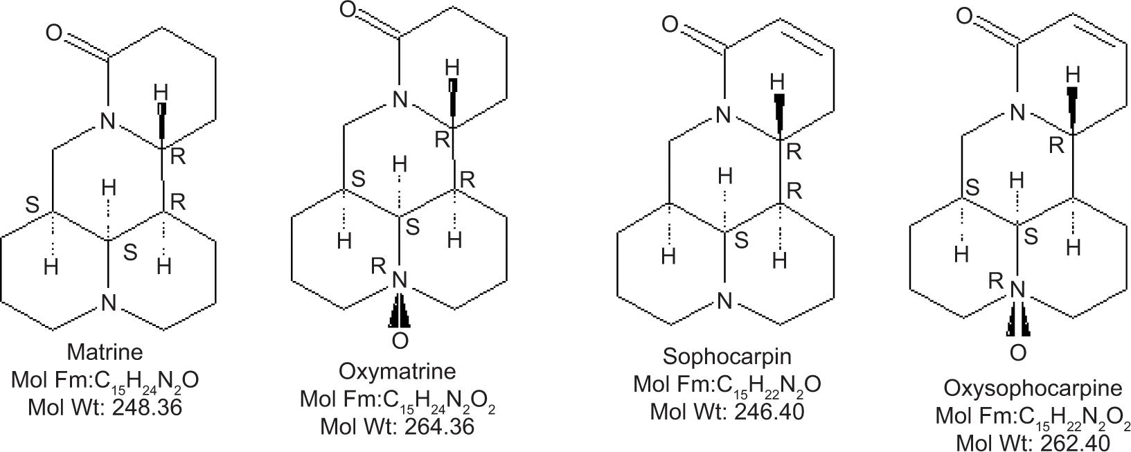
Figure S1 Chemical Structures of the four major alkaloids extracted from Kushen recipe.
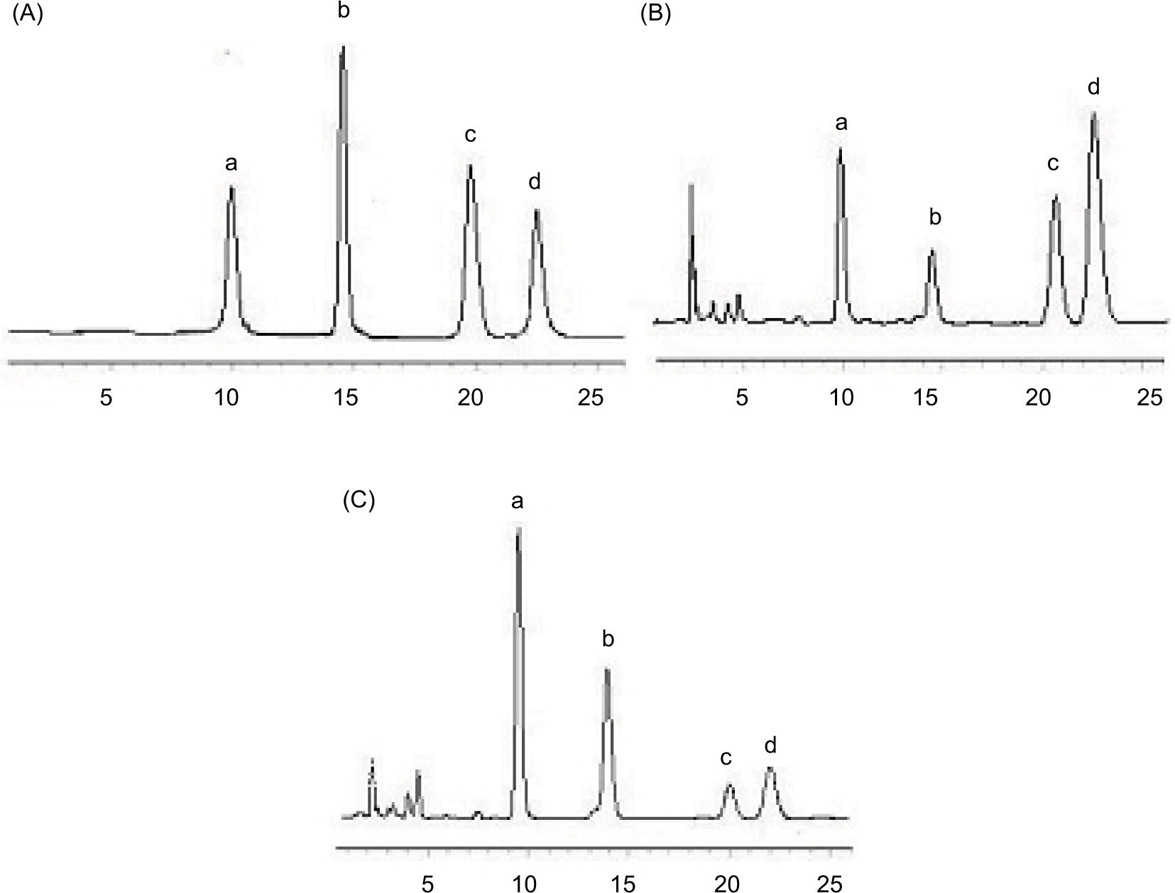
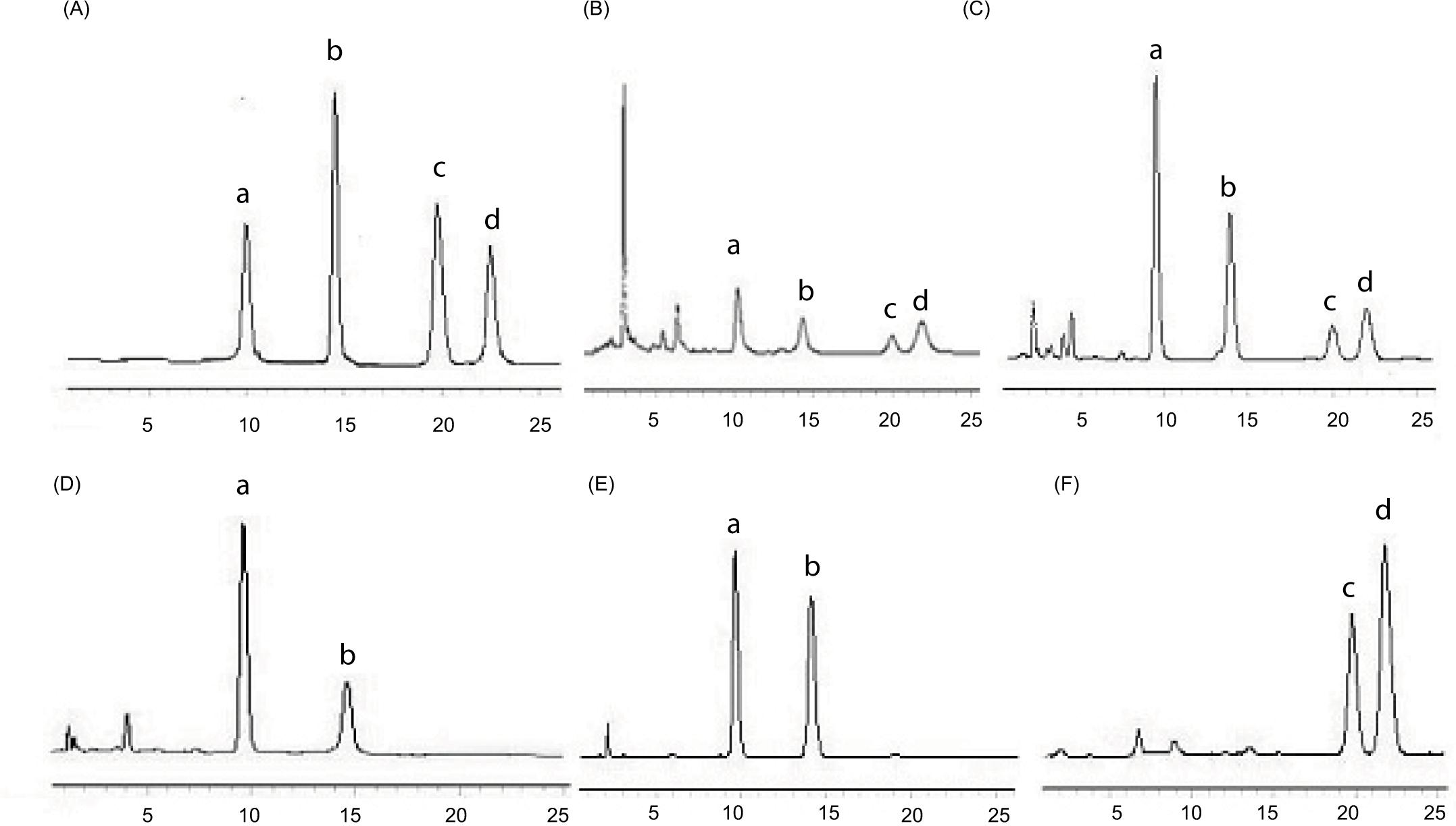
Figure S2 HPLC chromatogram. (A) Standard preparation, (B) Extract of Radix Sophorae Flavescentis, (C) Extract of Herba Schizonepetae and Radix Sophorae Flavescentis. a: Matrine, b: Sophocarpine, c: Oxysophocarpine, d: Oxymatrine.
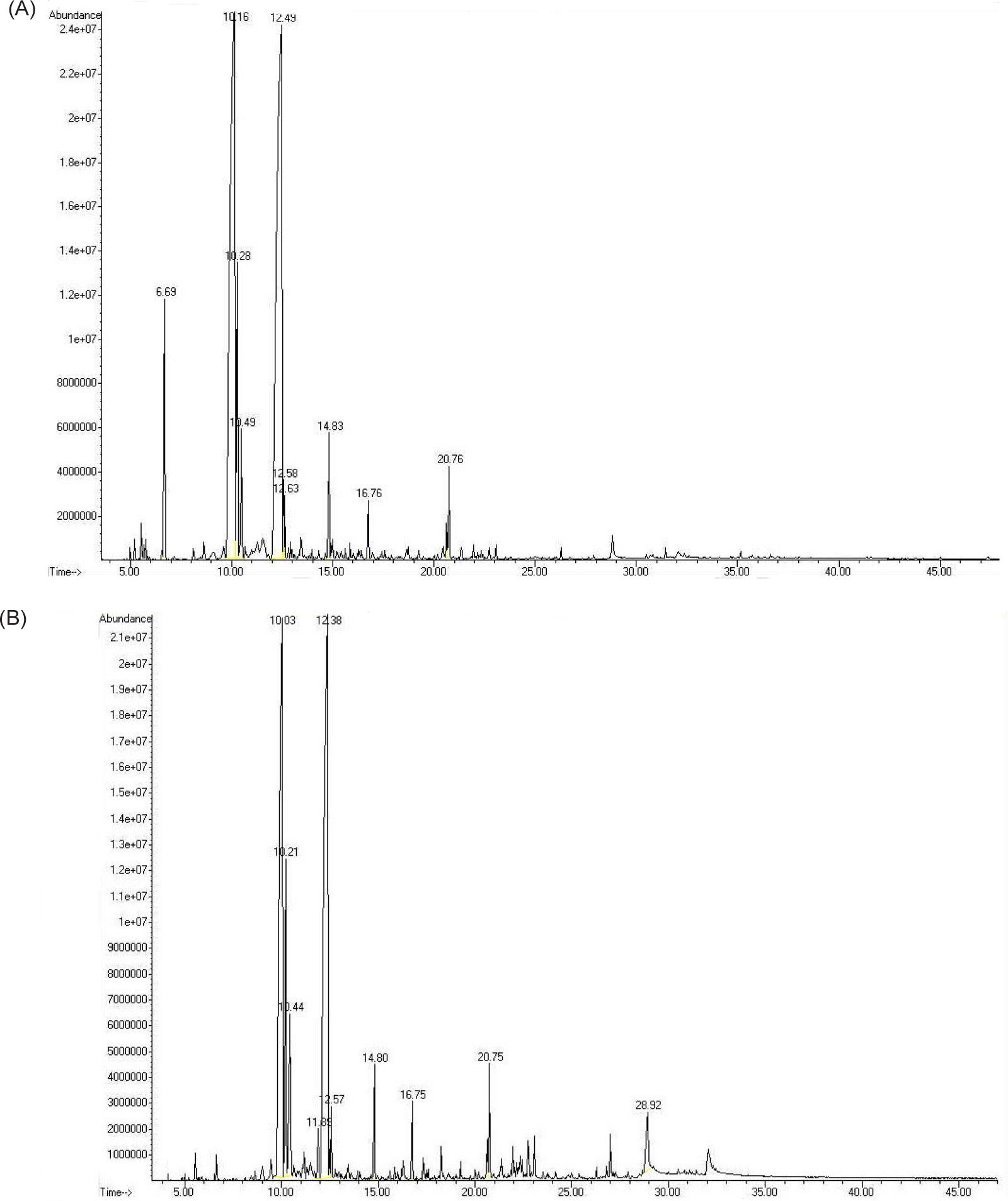
Figure S3 GC-MS total ion-current spectrum. (A) Volatile oil of Herba Schizonepetae, (B) Volatile oil of Herba Schizonepetae and Radix Sophorae Flavescentis.
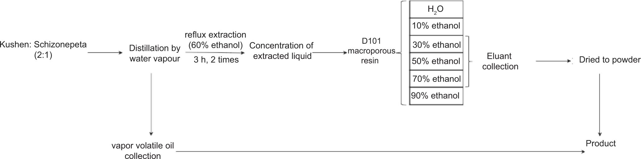
Figure S4 Extraction procedure of Kushen recipe active components.
Figure S5 HPLC chromatograms of eluents. (A) Standard substances, (B) Decotion of Kushen recipe, (C) Purifying extract of Kushen recipe. (D) Eluent of 30% alcohol, (E) Eluent of 50% alcohol, (F) Eluent of 70% alcohol. a: Mat, b: Sop, c: Osp, d: Omt.
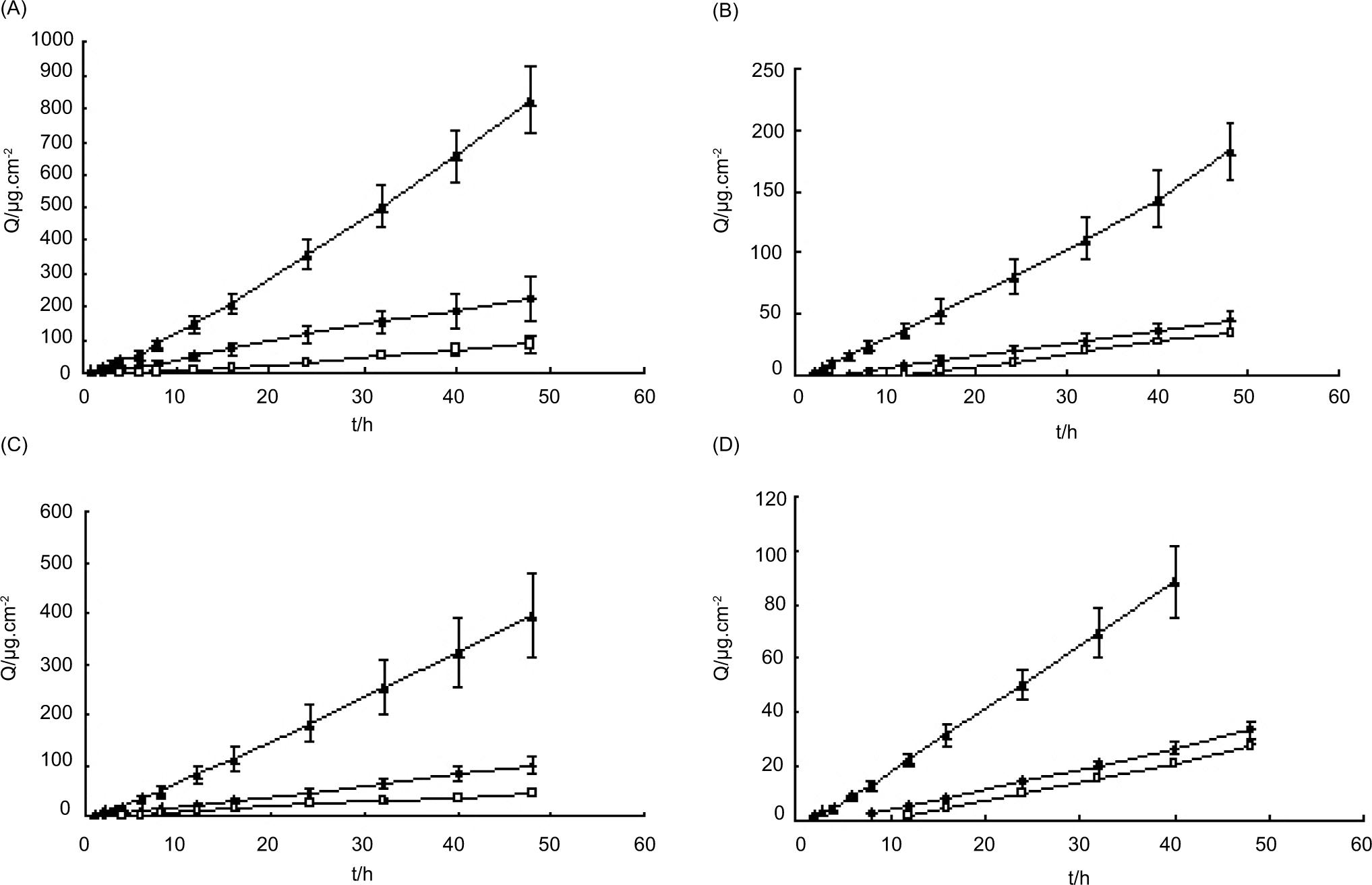
Figure S6 Cutaneous permeation curves of the four alkaloids of Kushen recipe (n=6). (A) Mat, (B) Sop, (C) Osp, (D) Omt. □ Decoction, ♦ Water solution of the extract, ▲ Kushen recipe gel.