
Download
CASE REPORT
Drug-induced fixed urticaria because of hypersensitivity to non steroidal anti-inflammatory drugs
Julia Jaque Gómez-Aguadoa,c*, Alberto Marín Muñoza, Beatriz Castillo Gómezb, Luis Moral Gilb,c
aPediatric Department, Dr Balmis General University Hospital, Alicante, Spain
bPediatric Allergy and Respiratory Unit, Dr Balmis General University Hospital, Alicante, Spain
cAlicante Institute for Health and Biomedical Research (ISABIAL), Alicante, Spain
Abstract
Drug-induced fixed urticaria (DIFU) is a rare skin reaction, characteri zed by pruritic hives that reappear in the same location after exposure to the triggering drug. In the few cases reported in the literature, it is considered as an atypical variant of fixed drug eruption (FDE). We present the case of a 10-year-old girl with allergic rhinoconjunctivitis, who had experienced three episodes of localized urticaria on the dorsal surface of both arms, 1 hour after taking ibuprofen. These manifestations did not appear when taking paracetamol, which she tolerated. Her father suffers from generalized urticaria to NSAIDs. An oral provocation test (OPT) was performed with ibuprofen, acetylsalicylic acid, and metamizole, which also caused the same fixed-location wheals and subsequent spontaneous resolution. She, however, tolerated an OPT with meloxicam. This case is notable because of the rarity of the DIFU and the familial aggregation of hypersensitivity to NSAIDs. In this case, as in those of DIFU described in the literature, the hives appeared shortly after the drug was administered and resolved rapidly. This feature distinguishes it from FDE, and it resembles acute drug urticaria, as in the patient’s father, which raises questions concerning the pathophysiology of DIFU.
Key words: NSAID hypersensitivity, fixed drug eruption, familial aggregation, pediatrics, drug-induced fixed urticarial
*Corresponding author: Julia Jaque Gómez-Aguado, Pediatric Department, Dr Balmis General University Hospital, Alicante, Spain. Email address: [email protected]
Received 1 July 2025; Accepted 10 September 2025; Available online 1 November 2025
Copyright: Gómez-Aguado JJ, et al.
This open access article is licensed under Creative Commons Attribution 4.0 International (CC BY 4.0). http://creativecommons.org/licenses/by/4.0/
Introduction
Drug-induced fixed urticaria (DIFU) is an immediate-type reaction, characterized by isolated pruritic wheals that always appear in the same site after exposure to the responsible drug. DIFU was first described by Barbarroja-Escudero and colleague s in 2015 in two adult patients with atypical fixed drug eruption (FDE), caused by local anesthetics, and by paracetamol and metamizole, respectively. A suspected IgE-mediated mechanism was considered by the results of skin biopsies in both of them.1 Another two adult patients with DIFU because of cross-reactive NSAID intolerance were described in 2019, being considered as a type of FDE that resembled a local form of NSAID-induced urticaria/angioedema (NIUA).2 A fifth case in an adult caused by nivolumab has just been newly reported.3 We are aware of only two recently published p ediatric cases, caused by NSAID and by plasma-derived activated prothrombin complex concentrate, respectively.4,5 We are reporting a third pediatric case (eight in adults and children) that presented at our center.
Clinical Case
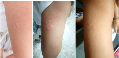
This case concerns a 10-year-old girl who reported three episodes of localized urticaria on the dorsal side of both upper arms (Figure 1) and on the eyelid in the last 2–3 months, which occurred approximately 1 hour after taking ibuprofen. The wheals faded without treatment after 1 hour, although they did reappear with less intensity in the subsequent hours. She had previously taken ibuprofen, paracetamol, and metamizole with no adverse reaction. Her medical history included allergic rhinitis with sensiti zation to house dust mites. Her father also suffers hypersensitivity to NSAIDs, presenting with urticaria on his arms and other sites, as well as eyelid edema after taking ibuprofen, acetylsalicylic acid, and metamizole, but with good tolerance to paracetamol.
Table 1 Characteristics of published patients with drug-induced fixed urticaria.
| Case | 1 | 2 | 3 | 4 | 5 | 6 | 7 | 8 |
|---|---|---|---|---|---|---|---|---|
| Age | 30 years | 50 years | 48 years | 50 years | 16 years | 10 years | 51 years | 10 years |
| Sex | Female | Female | Female | Female | Male | Male | Female | Female |
| Reference | 1 | 1 | 2 | 2 | 5 | 4 | 3 | Actual |
| Atopic or other diseases | Allergic rhinoconjunctivitis | Psoriatic arthritis | Hemophilia A with inhibitorFVIII | Allergic rhinitis | Metastatic melanoma | Allergic rhinitis | ||
| Culprit drug(s) | Local anesthetics: mepivacaine, lidocaine | Paracetamol, metamizole | Ibuprofen | Ibuprofen, dexketoprofen, ASA, metamizole, diclofenac | Plasma-derivedactivated prothrombin complex concentrate | Ibuprofen and ASA | Nivolumab | Ibuprofen |
| Time to reaction | 20 minutes | 15–20 minutes | 5–50 minutes | 30 minutes | Sudden onset | 20–60 minutes | 30 minutes | 1–3 hours |
| Time to spontaneous resolution | 15–30 minutes | 30 minutes | Less than 6 hours | 2 hours | 3 hours | 1 hour, but may flare up again mildly some hours later | ||
| Lesion location | Outer face of her right arm | Upper abdomen (epigastrium) | Right superciliary region | Rightsuperciliary region | Both antecubital regions | Both armpits | Décolleté | Dorsal side of both upper arms. Occasionally eyelid wheal |
| Allergy tests | Skin prick and intradermal tests negative to mepivacaine. Patch test negative to mepivacaine and lidocaine | Patch test negative to paracetamol and metamizole | Family rejected patch test | Patch test positive to ibuprofen and negative to acetylsalicylic acid (ASA) | Prick, intradermal, and subcutaneous tests negative to nivolumab | |||
| Reproduced index lesion after provocation tests | Subcutaneous challenge test positive to mepivacaine | Open challenge test positive toparacetamol and to metamizole | Single-blind oral challenge test positive to ASA | Single-blind challenge test positive to diclofenac | Reappearance after open administration of the culprit drug in spite of antihistamine premedication | Oral provocation test (OPT) positive to ibuprofen | Reappearance after repeated administration of nivolumab | Oral provocation test positive to ibuprofen, ASA, and metamizole |
| Other tests | Subcutaneous challenge test negative to bupivacaine | Single-blind oral challenge tests negative to paracetamol, meloxicam, and celecoxib. Negative test to diverse physicalstimulations (scratching, heat, and cold) | Single-blind oral challenge tests negative to paracetamol, meloxicam, and celecoxib. Negative test to diverse physicalstimulations (scratching, heat, and cold) | OPT negative to paracetamol | Oral provocation test negative to meloxicam | |||
| Biopsy | Urticarial lesion | Neutrophilic-type urticarial pattern | Patient rejected | Patient rejected | No | No | Superficial perivascular dermatitis withdermal edema and eosinophils | No |
| Treatment | No | No | Cetirizine with resolution in 15 minutes | Antihistamines with resolution in less than 1 hour | Antihistamine premedication ineffective | No | No | No |
| Outcome | Tolerates other NSAIDs | Rapid desensitization protocol successful in spite of urticarial plaques in both antecubital regions resolved with hydroxyzine after 45 minutes | Completes 4cycles of nivolumab | Tolerates paracetamol | ||||
| Proposed mechanism | Type I hypersensitivityreaction | Type I hypersensitivityreaction | Type of fixed drug eruption (FDE)resembling a local form of NSAID-induced urticaria/angioedema (NIUA) | Type of FDE resembling a local form of NIUA | Atypical presentation of FDE | Atypical presentation of FDE | Localized memory T-cell-induced mast cell activation via non-IgE pathways | Atypical variant of FDE or peculiar form of acute drug-induced urticaria |
An oral provocation test (OPT) was performed with ibuprofen (1/10, 1/4, and 1/1 of the therapeutic dose in intervals of 1 hour; total cumulative dose 540 mg), which gave a positive result with the immediate appearance of a wheal on the left eyelid which disappeared and, 2 hours after administration, the appearance of itchy erythematous wheals on the dorsal surface of both upper arms that resolved spontaneously without treatment. Subsequently, OPT was conducted with acetylsalicylic acid (total cumulative dose 785 mg) and with metamizole (total cumulative dose 810 mg), which caused the appearance of the same wheals on the dorsal surface of her arms between 1 and 3 hours after administration, which resolved spontaneously. The wheals were completely similar to those occurring in previous episodes (Figure 1). No other skin manifestations or systemic symptoms were noted. The girl continues tolerating paracetamol, and also tolerated an OPT with meloxicam. Other laboratory tests or the performance of a biopsy were not considered useful nor convenient for this patient.
Figure 1 Three different episodes of urticaria from the patient. Wheals appear in the same location in both upper arms 1–3 hours after the intake of different NSAIDs. The wheals faded away without treatment in about an hour and flared up again mildly some hours later. Pictures captured and supplied by her mother.
Discussion
Our case adds to the rare reports of patients with DIFU. The eight published cases to date, including the one presented in this report, are summarized in Table 1. Common characteristics of these patients are the mild course with the absence of systemic symptoms, the short interval between drug administration and urticarial lesions, usually 1 hour or less, and fast resolution even without treatment, in normally less than 1 to a few hours. DIFU may be more frequent than supposed, if patients and doctors are not worried about such a mild disorder.
Cross-intolerance to NSAID was the main cause of DIFU in four of the eight patients. In our patient, the father suffers from generalized urticaria because of hypersensitivity to NSAIDs. Although this familial aggregation is not reported in other cases of DIFU, a genetic component has been reported in both FDE6 and NSAID hypersensitivity.7
Allergy skin tests are mostly useless. Provocation tests should be performed for confirmation because they are safe and produce mild urticarial lesions in the same location as the index reaction. Provocation tests are also useful to find alternative drugs for the patient.
The biopsy performed in three of the patients shows findings of typical urticaria. Some authors consider DIFU as an atypical variant of FDE, with which it shares the recurring location of the wheals in the same site after exposure to the drug involved. However, FDE is a late hypersensitivity reaction that usually presents as erythematous-violaceous wheals with well-defined edges that appear between hours to days after administration of the causative drug. The wheals subsequently heal leaving a residual post-inflammatory hyperpigmentation.6,8 In contrast, in all cases reported in the literature with DIFU, the typical wheals developed and faded rapidly, without leaving residual lesions nor skin pigmentation. Patients with DIFU seem to share characteristics that are halfway between FDE and acute drug urticaria, and are also similar to some rare cases of food-induced fixed eruption.9,10 Therefore, DIFU may be considered either as an atypical variant of FDE or as a peculiar form of acute drug-induced urticaria.
Although the treatment consists in avoiding the drug or drugs involved, repeated administration or desensiti zation have been performed when the drug was essential.3,5
Conclusions
DIFU is a mild disorder characterized by repeated episodes of wheals in the same location after the administration of the same drug(s). DIFU is frequently associated with NSAID hypersensitivity, although its underlying physiopathology and long-term outcomes remain unknown because of the scarce number of patients who have reported to date. Provocation tests are safe and necessary for diagnosis.
Disclosure
The authors declare no potential conflicts of interest with respect to research, authorship, and publication of this article.
Author’s Contribution
All authors contributed equally to this article.
Conflicts of Interest
None.
Funding
None.
REFERENCES
1 Barbarroja-Escudero J, Sanchez-Gonzalez MJ, Rodriguez-Rodriguez M, Antolin-Amerigo D, Vélez D, Medina-Exposito I, et al. Fixed drug urticaria: a report of two patients. Allergol. Int. 2015 Jan 64(1):101–3. 10.1016/j.alit.2014.07.007
2 Argiz L, Múgica MV, Vega F, Blanco C. Drug-i nduced fixed urticaria as a presentation of NSAID i ntolerance. J. Allergy Clin. Immunol. Pract. 2019 Apr 7(4):1306–7. 10.1016/j.jaip.2018.10.030
3 Gambichler T, Weyer-Fahlbusch SS, Dengler S, Schaller J, Susok L. Consistently reproducible fixed drug-induced urticaria in a patient with metastatic melanoma under immunotherapy. Acta. Derm. Venereol. 2025 May 20;105:adv43652. 10.2340/actadv.v105.43652
4 Gultekin TTK, Emeksiz ZS, Selmanoğlu A, Misirlioglu ED. Nonsteroidal anti-inflammatory drug-induced fixed urticaria: first pediatric case report. Ann Allergy Asthma Immunol. 2024 Jul 4;133(4):470–2. 10.1016/j.anai.2024.06.030
5 Çelik HI, Akay E, Emeksiz ZS, Işık M, Yaralı HN, Mısırlıoğlu ED. Pediatric hemophilia patient: successful desensitization for drug-induced fixed urticaria with prothrombin complex concentrate. Pediatr. Allergy I mmunol. 2024 Mar 1;35(3). 10.1111/pai.14105
6 Anderson HJ, Lee J.B. A Review of fixed drug eruption with a special focus on generalized bullous fixed drug eruption. Medicina 2021; 57(9):925. 10.3390/medicina57090925
7 Jurado-Escobar R, Doña I, Triano-Cornejo J, Perkins JR, Pérez-Sánchez N, Testera-Montes A, et al. Genetic variants in cytosolic phospholipase A2 associated with nonsteroidal anti-inflammatory drug-induced acute urticaria/angioedema. Front Pharmacol. 2021;12:667824. 10.3389/fphar.2021.667824
8 Mathieu A, de Grandmont M, Fernandes CL, Kechichian E. Triggers, clinical manifestations and assessment of paediatric fixed drug eruptions: a systematic review of the literature. Contact Dermatitis. 2024;90(4):343–9. 10.1111/cod.14500
9 Parker AL, Pinson ML, Wohltmann WE, Gomez R. Fixed food eruption caused by peanut and cashew: a case report and review of the literature. J. Allergy Clin. Immunol. Pract. 2015 Jan–Feb;3(1):119–22. 10.1016/j.jaip.2014.08.004
10 Vichyanond P, Tanticharoenwiwat P, Wongteerayanee C, Rutrakool N, Senavonge A, Jeekungwal N, et al. Localized abdominal urticaria: a distinct clinical phenotype of wheat allergy in young children. J. Allergy Clin. Immunol Pract. 2020 Nov-Dec 8(10):3650–3652.e1. 10.1016/j.jaip.2020.06.010